
and Related Links
- Light sources on page O6
- Stage micrometers, eyepiece reticles & calibration slides on page S3
- CCD cleaning swabs on page M8
ProSciTech is the largest supplier of dedicated microscope cameras in Australia and New Zealand. Our cameras are effective, competent, reliable and very economical to purchase by customers all over the world.
Cameras are provided complete with comprehensive software for recording and editing of still and movie images, image analyses and display. We supply cameras with a built‐in C‐mount, using a CMOS chip for normal light levels and CCD models for very low‐light situations. Eyepiece adapters are available for a small fee for users with binocular Microscopes.
You can pay a lot more for a lot less in microscope cameras!
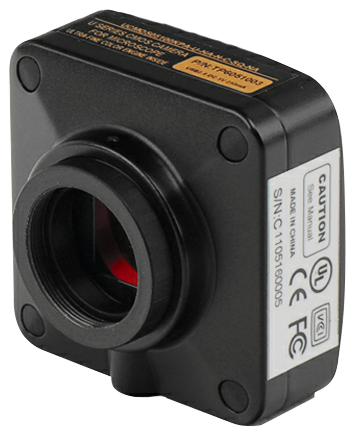

CMOS MICROSOPE CAMERA FOR C-MOUNT
USB2.0
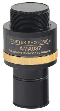
 Our high quality, affordable range of microscope digital cameras attach to any standard C‐Mount screw fitting. If you wish to place your camera through an available eyepiece port, we offer two types of standard eyepiece (23.2mm) adapters ‐ fixed and adjustable. As well as sleeves that slip into non‐standard eyepiece tube (30mm, 30.5mm). If you require the existing eyepiece to be par‐focal with the camera we suggest you purchase the adjustable adapter. If you have a third party microscope that does not provide a C‐Mount then please see the bottom of this page for a list of available adapters for some popular Nikon, Leica, Zeiss and Olympus microscopes.
Feel free to contact us about your set‐up and we'll be happy to help.
Cameras include a cable, CD with instructions and excellent, comprehensive software. These cameras use CMOS chips from the best Japanese and American manufacturers. Please be aware that the software included only runs on Windows (XP, Vista, 7), Mac OSX and Linux machines with an available USB 2.0 port.
Please note that the OSX version of the software does not have all of the additional features, eg measuring and calibration, that is included in the Windows version.
For some basic tutorials on how to use ToupView X, please visit the following youtube channel.
Subscribe to be notified when a new tutorial is uploaded!
NEW! Software now available for Mac OSX and Linux! If you have previously purchased a camera from us and would like a copy for your Mac or Linux machine please contact us.
SALE: The manufacturer gave us a big price‐break on all CMOS microscope cameras. These are the best selling cameras in Australia and New Zealand; they are now an even greater bargain and worth shipping to anywhere.


Documents


USB3.0 HIGH SPEEED
New Product! Just released from the manufacturer are these new USB3.0 microscope cameras. Using the same design as our USB2.0 cameras above, the USB3.0 cameras bring the benefit of added speed, up to ten times the data transfer speed of USB2.0. We see USB3.0 taking over from USB2.0 in the long term so you should consider investing in this new technology.
The USB3.0 cameras use the same eyepiece adapters available for the USB2.0 cameras above. The software used is the same, with a few tweaks to allow for the higher transfer rate. Because of the USB3.0 interface, the cameras are slightly larger in size, but the same style.
The majority of digital microscope cameras use a version of the USB interface to pass the image data to a computer. There was no official industry standard for using USB2.0 in imaging devices, however it was widely accepted. For some more specialised cameras, the GigE interface was used due to its higher speed and longer cable length. With the introduction of USB3.0 in 2008, it has gained popularity for imaging use due to its increased speed. At the end of 2012 the Automated Imaging Association (AIA) released a Vision Standard for USB3.0 in imaging devices. The GigE interface can still be found on cameras that require a cable length longer than approximately 5 metres where USB cabling is not ideal.
In terms of how this affects microscope cameras; the advantage of USB3.0 is a faster transfer speed that allows for a higher frame rate and for image data to be saved to disk quicker.
Before purchasing a USB3.0 camera, insure that your computer has an available port.
If you don't have access to the computer specifications, locate your USB ports and look for a blue accent on the inner prong.
We recommend consulting your IT department before making your purchase.
| Order Code | Sensor & Size(mm) | Pixel(μm) | G Sensitivity Dynamic Range SN Ratio | FPS/Resolution | Binning | Exposure |
|---|---|---|---|---|---|---|
O3HS1400 | 14M/MT9F002(C) 1/2.3" (6.451x4.603) | 1.4x1.4 | 0.724v/lux‐sec | 6.2@4096x3286 | 1x1,2x2,4x4 | 0.4ms~2000ms |
O3HS1000 | 10M/MT9J003(C) 1/2.3" (6.44x4.616) | 1.67x1.67 | 0.31v/lux‐sec | 7.2@3584x2746 | 1x1,2x2,4x4 | 0.38ms~2000ms |
O3HS0850 | 8.5M/Special(C) 1/2.5" (5.557x4.255) | 1.67x1.67 | 0.31v/lux‐sec | 8.3@3328x2548 | 1x1,2x2,4x4 | 0.1ms~2000ms |
O3HS0510 | 5.1M/MT9P006(C) 1/2.5" (5.7x4.28) | 2.2x2.2 | 1.76v/lux‐sec | 14.2@2560x1922 | 1x1,2x2,4x4 | 0.05ms~2000ms |
O3HS0320 | 3.1M/AR0330(C) 1/3" (4.505x3.38) | 2.2x2.2 | 1.9v/lux‐sec | 27.3@2048x1534 | 1x1,2x2 | 0.1ms~2000ms |
Available on request | 1.2M/AR0130(C)1/3"(4.8x3.6) | 3.75x3.75 | 5.5v/lux‐sec 85.3dB 44dB | 45@1280x960 55@640x480 | 1x1,2x2 | 0.1ms~20000ms |
| OTHER HARDWARE CONFIGURATION | |
|---|---|
Spectral Range | 380‐650nm (with IR‐filter) |
White Balance | ROI White Balance/ Manual Temp‐Tint Adjustment |
Color Rendering Technique | Ultra Fine Color Engine |
Capture/Control API | Native C/C++, C#, Directshow, Twain, Labview |
Recording System | Still Picture and Movie |
Cooling System* | Natural |
| OPERATING ENVIRONMENT | |
Operating Temperature | ‐10℃~ 50℃ |
Storage Temperature | ‐20℃~ 60℃ |
Operating Humidity | 30~80%RH |
Storage Humidity | 10~60%RH |
Power Supply | DC 5V over PC USB Port |
| SOFTWARE ENVIRONMENT | |
Operating System | Support Microsoft Windows XP / Vista / 7 /8 /10 (32 & 64 bit) |
PC Requirements | CPU: Equal to Intel Core2 2.8GHz or Higher |
Memory: 2GB or More | |
USB port: USB3.0 High‐speed Port | |
Display: 17" or Larger | |
CD‐ROM | |
stream live feed to wifi enabled computers or hand-held iOS and android devices
These cameras are similar to the CMOS cameras above however they generate a WiFi signal for sending high‐resolution stills and video from a microscope to a WiFi‐enabled device such as smartphones, tablets, and computers with iOS, Android, OS X, Linux and Windows operating systems, streaming images to up to six devices simultaneously.
The camera includes ToupView images software for quantifying, measuring, and annotating images and for using with an interactive white board. It also works with the free, downloadable Toupview app for viewing, capturing, and editing images.
Please note that a USB cable is still required to power the device, but can simply be plugged into a USB wall socket.
| Order Code | Sensor & Size(mm) | Pixel(μm) | G Sensitivity Dynamic Range SN Ratio | FPS/Resolution | Binning | Exposure |
|---|---|---|---|---|---|---|
OWCAM1080 | 1080P/IMX222(C) 1/2.8"(7.6x5.8) | 2.8x2.8 | 510m with 1/20s | 25@1920x1080 | 2x2 | 0.059ms~1941ms |
OWCAM0720 | MT9P001(C) 1/2.5"(5.70x4.28) | 2.2x2.2 | 1.0 V/lux‐sec | 30@1280x720 | 2x2 | 0.21ms~200ms |

(video mode)
CMOS chips have become most popular in battery operated cameras because they use one hundredth of the electricity compared with CCD chips. For a given size CCD sensors are considerably more light efficient than CMOS, and that makes these cameras particularly suitable for fluorescence microscopy. The 1.4MP camera's CCD has an active surface area of 57mm². If that area is divided by the number of pixels and similarly the area of the 3.2Mpixel camera is divided by its pixels, the surface area/ pixel is 4x larger on this low light camera. Accordingly, these larger pixels receive 4x as many electrons. That parameter makes the 1.4MP camera four times more sensitive. Additionally, this camera has a much longer exposure time available. These sensors are made by Sony, and the extraordinary 1.4MP is among the most light-efficient un-cooled chips available. They are made to order requiring 2 months delivery. These lights are great for fluorescence microscopy. This camera can be supplied within one month.
| Hardware Information | |
| Specifications | 1.4MP CCD |
| Scan Mode | Progressive |
| Max resolution | 1360 x 1024 |
| Image sensor size | ²/3" (16.93mm) |
| Pixel size | 6.45µm x 6.45µm |
| G Sensitivity | 1240mV with 1/30s accumulation |
| Dynamic Range | 70dB |
| A/D Converter | 12-bit parallel, 8-bit R.G.B. to PC |
| SN Ratio | 62dB |
| Spectral Range | 400 - 650nm (with IR filter) |
| Frame Size/Rate | 15fps @ 1360 x 1024 |
| Binning | 1 x 1 |
| Exposure | Normal: 0.126 ~ 66.695ms Long: 66.695ms ~ 4 mins |
| Colour Rendering Technique | Ultra Fine™ Colour Engine |
| White Balance | One push ROI / manual temp-tint adjustment |
| Capture/Control API | DirectShow / Twain |
| Capture Mode | Still picture and video |
If you require an eyepiece adapter for this camera, please see the adapters above in the CMOS cameras section.
We suggest; ODCMZ-EAF-7, fixed 0.75x magnification lens.

Testimonial
Testimonial from one of Australia's large University Microscopy Centres - they bought a 3.2 megapixel eyepiece camera:
"We have now tried the camera and as you predicted, we are very pleased with it. It is better than the XXX top-brand camera we had on there previously, that cost 10 times as much and they now want $1500 to repair".


SOME COMMENTS ON DIGITAL MICROPHOTOGRAPHY
Since the advent of digital cameras, micrography has become much easier. The temptation is to use a conventional digital camera with many mega-pixels of image capacity and adapt that to the microscope. It is possible, but few such cameras are well suited to microscopy.
Photography is easier when the camera is connected to microscope and computer to display the microscope's view continuously on the computer screen: you see what you get, and focus on the large screen-image. Cameras with an attached very low power eyepiece are very easy to use, economical to purchase and give best results. C-mounts work equally well, but the C-mount is an additional purchase and the camera may not as easily changed to another microscope.
It must also be recognised that large image formats (pixel) are not often an advantage to the microscopist. The microscope's optics and not 'pixel' should be used for most of the magnification required. Camera magnification in micrography is kept low through the use of a low magnification eyepiece, since the small format of the CCD or CMOS (when compared with that of film cameras) results in larger magnifications - by ratio. In the upper magnification range of a compound microscope it is easy to display and photograph images at 10,000x - giving large, but very blurry images! Compound microscopes are naturally limited to ~ 1,000x and those higher powers are useless.
Enlargements of low and medium power images of suitable specimens are possible with multi-mega pixel cameras. However, most published micrographs had little photographic enlargement and could have been captured with our small 1.3m pixel camera; however, there are some situations when enlarging and therefore a larger (pixel) camera is beneficial. We offer a range of cameras for microscopy from 350K pixel to 9M pixel. The smaller the diagonal of the chip (CCD) the lower the eyepiece's magnification must be.![]() All items on this page are CE certified.
All items on this page are CE certified.


CMOS MICROSCOPE CAMERAS
For use with computers with Windows XP, Windows Vista or Windows 7 operating system. These easy to use and very versatile digital cameras are supplied integral with a low power microscope eyepiece (23.2mm DIN standard). They are simply inserted into an eyepiece tube or top tube of a trinocular head. The visual field observed in the camera is up to 90% of the viewing field through the eyepiece. Using the USB2.0 high speed port the camera displays the microscope images continuously on the computer screen in real-time and non-compressed video data and digital photos and movie sequences may be captured.
Prior to capturing an image, all adjustments - colour, contrast, brightness frame-speed - may be optimised. After capture, image files are saved in the desired image-format on the computer's hard drive. Once calibrated using a stage micrometer, the software allows measurement of lengths, angles and areas across microscope images. It is also possible to include a scale marker on micrographs and to count particles. For counting select Plugin and then Count, after that set parameters and Count.
Our cameras provide high image quality, and they are entirely suitable for numerous applications in research, in biomedical applications, material science and technology. The CCD camera has a "Wide Lux" feature. This largely compensates for changes in brightness that are due to changes in magnification and diaphragm aperture. These CCD cameras also have a "High Sensitivity" feature to compensate for a wide range of light-attenuating accessories, such as a dark-field" condenser, phase contrast and epi-fluorescence microscopy.
All of these cameras come with USB2.0 cable, driver software and sleeve adapters for inserting the camera into 30mm or 30.5mm tubes, which are commonly used with stereo microscopes.
Connecting a camera to the microscope: digital cameras for microscopy are usually connected to a microscope by one of two means: Cameras with an integral eyepiece are inserted into the phototube of a trinocular microscope (ready to receive an eyepiece), or they can be inserted into one of the viewing eyepiece tubes after an eyepiece is removed.
Alternatively the microscope has a C-mount adapter in lieu of the phototube. The C-mount camera has a thread which connects to the top of the C-mount adapter.
Photo magnification is affected not only by the microscope and C-mount/ camera eyepiece, but also by the camera chip and the size of the computer screen. Most microscopists would aim for a magnification which is larger, but less than double the viewing magnification. Compared with the settings for the viewed magnifications, our cameras, using a 0.5x C-mount/ eyepiece and displayed on a 22" screen, average magnifications 1.9x greater in full screen mode and 1.4x in working mode.
Particularly using macro photography or stereoscopes, a 1x C-mount is another means to obtain higher magnifications. A 1x C-mount used with a compound microscope and oil-immersion, results in several thousand times magnifications. Conventional microscopy is limited by the wavelength of light; magnifications above 1000x are increasingly blurred, and magnifications above 2000x have no merit. A 0.3x C-mount obviously results in lower magnifications, which can be useful, but in some systems may cause vignetting.
| Hardware Information | |||||
| Specifications | ODCM0130C | ODCM0310C | ODCM0510C | ODCM0900C | ODCM1400C |
| Pixels | 1.3MP (Approx.1,300,000 pixels) | 3MP (Approx.3,200,000 pixels) | 5MP (Approx.5,000,000 pixels) | 9MP (Approx.9,000,000 pixels) | 14MP (Approx.14,000,000 pixels) |
| Max resolution (video mode) | 1280 x 1024 pixels | 2048 x 1536 pixels | 2592 x 1944 pixels | 3488 x 2616 pixels | 4096 x 3288 pixels |
| Image sensor | ¹?3" CMOS chip, colour 8.46mm | ½" CMOS chip, colour (Diagonal 8.19mm) | ¹?2.5" CMOS chip, colour (Diagonal 7.13mm) | ¹?2.4" CMOS chip, colour (Diagonal 7.281mm) | ¹?2.3" CMOS chip, colour (Diagonal 7.672mm) |
| Preview speed/ Recording speed | 1280 x 1024 15fps, 640 x 512 26fps, 320 x 256 50fps | 2048 x 1536 8fps, 1024 x 768 22fps, 680 x 510 43fps | 2592 x 1944 5fps, 1280 x 960 18fps, 640 x 480 60fps | 3488 x 2616 2fps, 1744 x 1308 8fps, 872 x 654 27fps | 4096 x 3288 1.8fps, 2048 x 1644 10fps, 1024 x 822 27fps |
| Imaging area | 4.6mm(H) x 3.7mm(V) | 6.55mm x 4.92mm | 5.70mm x 4.28mm | 5.825mm(H) x 4.369mm(V) | 6.138mm(H) x 4.603mm(V) |
| Pixel size | 3.6μm x 3.6μm | 3.2µm x 3.2µm | 2.2µm x 2.2µm | 1.67µm x 1.67µm | 1.4μm x 1.4μm |
| Recording System | Still picture and video | ||||
| Master Clock - Hori. drive frequency | 54MHz | 48MHz | 54MHz | 96MHz | |
| A/D | 10-Bit parallel, 8-Bit R.G.B | 10-Bit parallel, 8-Bit R.G.B | 12-Bit on-chip, 8-Bit R.G.B | 12-Bit on-chip, 8-Bit R.G.B | 12-Bit on-chip, 8-Bit R.G.B |
| Binning | 1x1, 2x2 , 4x4 | 1x1, 2x2, 3x3 | 1x1, 2x2, 4x4 | 1x1, 2x2, 4x4 | 1x1, 2x2, 4x4 |
| Peak Quantum Efficiency | 30% | ||||
| Dynamic range | 71dB | 61dB | 66.5dB | 65.2dB | 65.3dB |
| Sensitivity | 1.0v/lux-sec @550nm | 1.0v/lux-sec @550nm | 0.53v/lux-sec @550nm | 0.33v/lux-sec @550nm | 0.724v/lux-sec @550nm |
| Spectral Range (Range of wavelength) | 400-650nm (with IR cut) | ||||
| Exposure Time | 0.142ms-2000ms | 0.128ms-2000ms | 0.21ms-2000ms | 0.38ms-2000ms | 0.4~2000ms |
Connecting mode:
Inserts directly into eyepiece tube. Eyepiece 23.2mm - standard DIN, two adaptors are included to fit 30mm, 30.5mm stereo microscope eyepiece tubes.
Specifications:
Description: Black metal cylinder body
Carton package: 145mm (L) x 145mm (W) x 90mm (H)
Weight(w/ USB cable): 280 g
Gross weight: 520 g
Lens Cover: Ø23.2mm rubber cover
Adaptors: Ø23.2mm to Ø30.0mm, Ø23.2mm to Ø30.5mm
Optic system: CFS anti-dust achromatism system
Filter: 400-650nm IR-cuter (380-1000nm w/o IR-Filter - ask)
Fitted with anti-blooming protection
DSP (Digital signal processing): Ultra fine™ colour engine
Optical input window: Fused silica
Reduction Lens: 0.5x relay lens to Ø23.2mm eyepiece (camera)
User manual
Digital cameras without the infra red filter are available - ask
Operating Environment:
Operating Temperature: 0°C to 40°C
Storage Temperature: -20°C to 60°C
Operating Humidity: 30%~80%
Storage Humidity: 10%~60%
Power Supply: PC USB2.0 high speed port [DC 5V]
Software Information:
Interface: USB, hot plug and thrust, USB cable length 1.5m
Software: Driver, ToupView image processing software
Driver interface support: DirectShow, Twain
Image download: USB2.0
File format: JPEG, PNG, TIF, PCX, TGA, SFT, PSD, etc.
System requirements:
- OS: Microsoft® Windows® XP / Vista / 7 / 8 (32bit or 64bit), OSX 10 >, Linux
- USB Port: USB2.0 high speed port
- CPU: Equal to Inter Core2 2.8GHz or higher
- RAM: 2GB or more memory
Documents


FINAL MAGNIFICATION
Final magnification in microscopy depends on several factors. In stereo or macro systems the magnification at the eye is the magnification of the objective multiplied by that of the eyepiece. If an additional lens is added to the front of the objective, both, photo and viewing magnifications, whereas the use of different eyepieces changes viewed magnifications only. Photo magnification is subject to other, additional factors.
Photographic magnification is unaffected by the viewing eyepiece, as the eyepiece is either substituted with the camera or optically bypassed, depending on the system. The optical magnification of C-mount or eyepiece cameras changes final magnification, but the mount magnification is usually selected to suit the format of the camera and obtain on the computer screen a magnification similar as seen through the eyepiece.
It is useful to know what the normal focal length lens of your camera format is. This is the diagonal of the sensitive (historically film) area. In the 35mm film format, to which we frequently relate the focal lengths of digital cameras, the diagonal is 43.3mm. With a little extra for coverage and to avoid vignetting, 45-55mm lenses are considered normal. 1/3" sensors have a normal focal length of 6mm, for ½" it is 8mm and a 2/3 sensor it's 11mm. In digital cameras the diagonal of the sensor is more than the lenses accepted as normal. Normal lenses render images with a viewing angle of around 53°. Most of our CMOS cameras have a chip close to a ½".
Approximately "normal" magnification at the screen depends on the size of the viewing monitor, and especially the CMOS/ CCD size and the eyepiece (or C-mount) magnification. The smaller the sensitive area, the lower the eyepiece needs to be, otherwise it is easily possible to achieve low resolution images from a light microscope with apparent magnifications of several thousand times. Full frame (35mm) cameras require an 8x eyepiece. A large CMOS microscopy camera works best with a 1x eyepiece, but a 12.7 or a 8.4mm lens requires a 0.5 times eyepiece or C-mount. Because magnification changes with enlargement (monitor size too), micrometer or mm bars are practical in micrography - see our link ToupView™ - Guide to calibration, measuring & counting

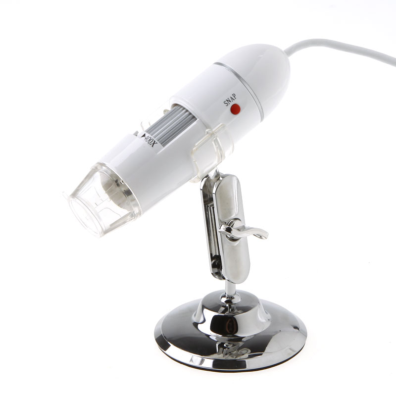
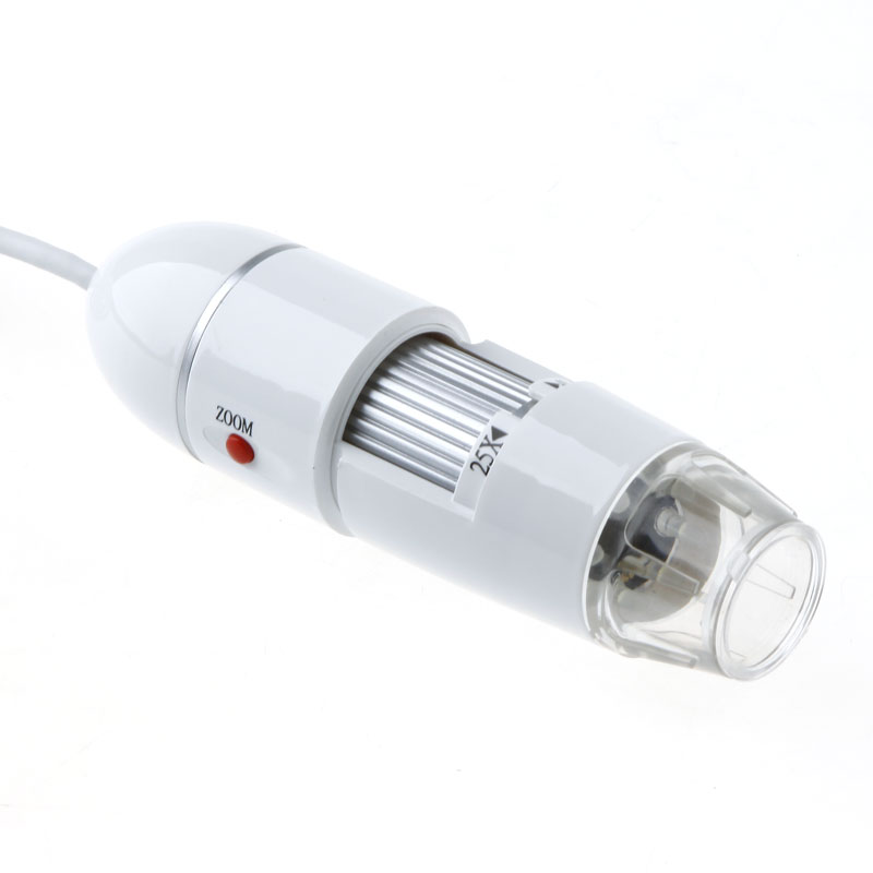
Digital magnifier

This is the high quality electronic microscope instead of the normal microscope. You can use it to snap HD pictures and capture the video, and also to measure the objects.
You can use this microscope for: Industrial Inspection, Computer Parts inspection, Telecom module inspection, scientific teaching tool, Laboratory Research, Medical analysis, School Research tool, Insect dissection examination, Plant dissection examination, Skin examination, Scalp examination, Textile Inspection, Jewelry Inspection, Collections/Coin Inspection, Printing Inspection, Reading Aid, Teacher Add as Project, Presentation and so on.
This inspection camera has many applications including industrial inspection, electronic accessories inspection, plant dissection/examination, skin examination, textile inspection, jewelry inspection, collections/coin Inspection, printing inspection, inspection dissection/examination, PCB or PCBA inspection and so on.
Features
- Mage HD Colour CMOS Sensor
- High Speed DSP Controller
- Micro‐Scope Lens
- 5X digital zoom
- Magnification adjustable ‐ Manual Focus range from 3mm to 40mm
- Capture snapshot image or video.
- It has 8 built‐in LED lights on camera head to illuminate the inspection area. The LED lights' brightness is adjustable.
- A alloy stand to support the camera.
- User Manual is included in the CD.
- Digital Measurement software and calibration ruler.
Specifications
| Cat. No. | ODMU400XW | ODMU500XB |
| Colour | White | Black |
| Still image and video capture resolution | 1600 x 1200 (2M Pixel) 1280 x 960 (1.3M Pixel) 800 x 600 640 x 480 | |
| Frame rate | Max. 30f/s Under 600 Lux Brightness | |
| Flicker control | 50Hz / 60Hz Option | |
| Controller | 24Bit DSP | |
| Magnification | 25x ‐ 400x (Manual) | 50x ‐ 500x (Manual) |
| Still image format | JPEG | |
| Video format | AVI | |
| LED light | 8 White Light LED (with controller on USB cable) | |
| Interface | USB2.0 & USB1.1 | |
| Product size (diameter x length) | 32 x 114mm | |
| Item weight | 80g | |
| Package size | 230 x 135 x 60mm | |
| Package weight | 300g | |
| Powered | USB Port (5V DC) | |
System Requirements
- Pentium Computer with 700MHz & Above
- 20M HD Space CD ROM Driver
- 128MB RAM
- Direct x VGA Card
- Operation System: Windows 7 32 bit /Vista/XP
Package includes:
1 x USB Microscope
1 x Plastic stage micrometer / emandation rule
1 x Alloy Stand Cover
1 x CD Software AMCAP (Driver)


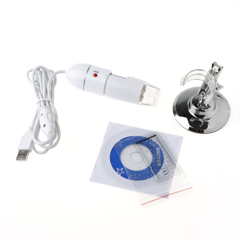

Documents
N/A
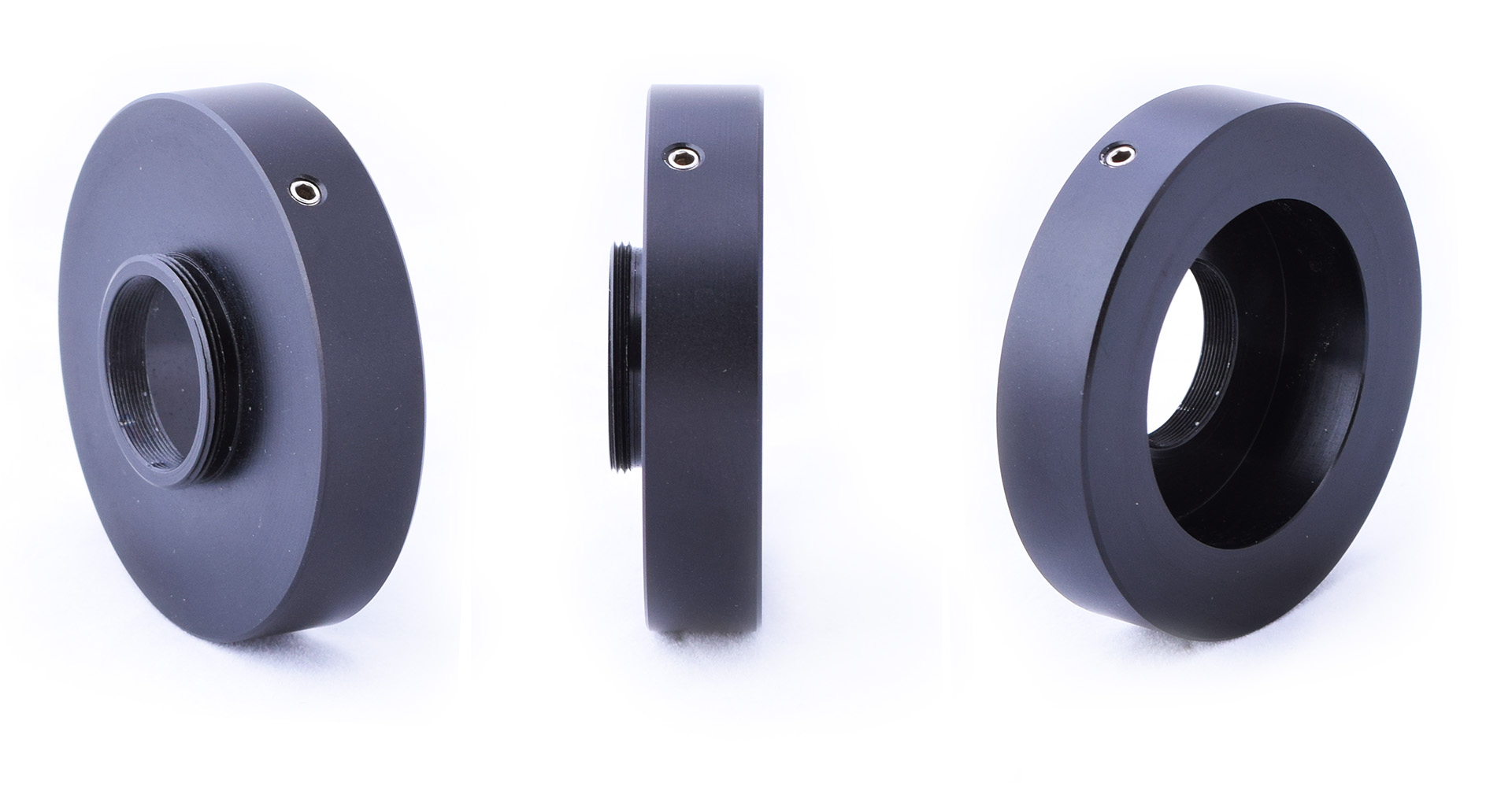
Cat. No. | Mag. | Style | Cmount | Trinocular Attachment | Overall Dimensions | Description | ONKCC01 | TV Tube | Diameter: 38mm Height: 7.7mm Double lip for screw | Diameter: 52-59mm (taper) Height: 84.4mm | TV tube takes 38mm C-mount adapters in the top, and connects to a 38mm microscope trinocular port. Used to extend focal distance. | ONKCC02 | 0.55X | MBF550 | 1'x1/32 | Diameter: 38mm Height: 13mm Indent for screw | Diameter: 50mm Height: 30mm | A simple C-mount attachment. Drops into 38mm TV tube (ONKCC01), or direct into microscope and tightens into an indent. Rotatable. | ONKCC03 | 1X | 1'x1/32 | Diameter: 38mm Height: 22mm Indent for screw | Diameter: 51mm Height: 38mm | A simple C-mount attachment. Drops into 38mm TV tube (ONKCC01), or direct into microscope and tightens into an indent. Rotatable. | ONKCC04 | 0.63X | 1'x1/32 | Diameter: 38mm Height: 25.3mm Indent for screw | Diameter: 51mm Height: 30mm | A simple C-mount attachment. Drops into 38mm TV tube (ONKCC01), or direct into microscope and tightens into an indent. Rotatable. | ONKCC05 | 0.55x | 1"x1/32 | Diameter: 40mm Height: 13mm Indent for screw | Diameter: 50mm Height: 30mm | A simple C-mount attachment. Drops into 38mm TV tube (ONKCC01), or direct into microscope and tightens into an indent. Rotatable. |


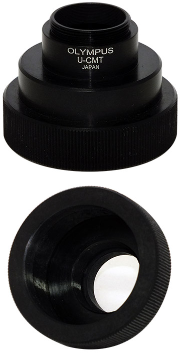
Cat. No. | Mag. | Style | Cmount | Trinocular Attachment | Overall Dimensions | Description | OYCC01 | U-CMAD3 U-TV-2 | 1"x1/32 | Diameter: 42mm Height: 6mm Tapered for screw | Diameter: 65mm Height: 93mm | Simple C-mount adapter. Drops into a 42mm trinocular opening and tightens into a tapered section. Rotatable. Non-adjustable. | OYCC02 | BH2 | 1"x1/32 | Fits over a 50mm trinocular 3 screw down points 30mm apart | Diameter: 58mm Height: 43mm | The underneath of this adapter has three tiers. The first is 35mm in diameter, the second is 50mm at which point 3 screw in points should be available on the microscope to secure the adapter to the body, there is a final tire that is 54mm. There should be an indent in the top of your microscope to allow for this final tier and for the adapter to sit flush. *Bolts not included* Non-rotatable. Non-adjustable. | OYCC03 | U-CMT | 1"x1/32 | Screws onto a 41mm thread | Diameter: 55mm Height: 41.5mm | Screws directly onto a 41mm thread. Would allow height adjustment via screw thread for about 20mm. | OYCC04 | 0.5X | Lock-Focus | 1"x1/32 | Diameter: 42mm Height: 6mm Tapered for screw | Diameter: 60mm Height: 42mm | Adjust focus using the screw on the left, then fix that position using the screw on the right. Rotatable. | OYCC05 | 0.63X | Lock-Focus | 1"x1/32 | Adjust focus using the screw on the left, then fix that position using the screw on the right. Rotatable. | OYCC06 | 1x | 1"x1/32 | Fits over a 38mm trinocular one screw down point | Diameter: 59mm Height: 16.5mm |


1x
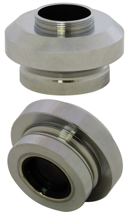
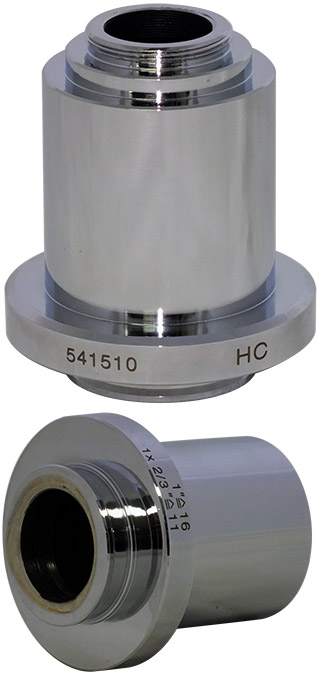
Cat. No. | Mag. | Style | Cmount | Trinocular Attachment | Overall Dimensions | Description | OLCCC01 | 1"x1/32 | Diameter: 34mm Height: 8mm Bottom lip for screw | Diameter: 54mm Height: 39mm | OLCCC02 | 541510 HC | 1"x1/32 | Diameter: 34mm Height: 7.3mm Bottom lip for screw | Diameter: 54.1mm Height: 63mm | Made from polished stainless steel. Very solid construction. |

1x
Cat. No. | Mag. | Style | C-mount | Trinocular Attachment | Overall Dimensions | OCZCC02 | 45610501 60C | 1"x2/3 | Diameter: 30mm Height: 10.9mm Indented for screw | Diameter: 46.9mm Height: 57.8mm | OCZCC03 | 45 6006 | 1"x2/3 | Turns trinocular into third eyepiece port (30mm inner diameter). |


CAMERA ADAPTERS, C-MOUNT TO DSLR
Our cameras come with image processing software, ToupView™, this includes some analysis functions like particle counting and measurements. However, if you want more elaborate image analysis software, these software packages maybe suitable.